University of Minnesota Scientists Find New Diagnostic Signatures and Therapeutic Targets in Gene Expression Data
by Wes Conard
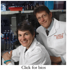 Wife and husband team, Drs. Amy and Keith Skubitz, discuss the challenges of expression analysis in ovarian cancer, renal cancer, and sarcomas Amy and Keith Skubitz are engaged in two major collaborations that vie for literally every minute of their time. The first project involves a small group of cohorts— their four young children—set against the variables of school, soccer practice, playtime and homework. The second collaboration, finding new ways to diagnose and treat a wide range of cancers, gets most of their remaining attention, once the school bus leaves in the morning and after the kids fall asleep at night. Dr. Keith Skubitz is a professor of medicine in the hematology, oncology, and transplantation division at the University of Minnesota and has studied gene expression of soft tissue cancers and renal carcinoma for the past half decade. “We really have two problems when it comes to diagnosing cancers like soft-tissue tumors,” said Keith Skubitz. “One, within a diagnostic category there's really more than one entity. And two, even the reproducibility of how we currently put samples into these categories is not great.” In a recent Journal of Clinical Laboratory Medicine publication, Keith Skubitz led the research that used microarray-based gene expression profiling to classify high grade pleomorphic sarcomas and malignant fibrous histiocytomas with known entities—results that were ultimately confirmed in several instances through more detailed pathological and biochemical exams. Dr. Amy Skubitz, an associate professor in the department of laboratory medicine and pathology at the same institution focuses her research on developing new methods to detect ovarian cancer, a disease that is 90 percent curable when detected early, but is currently the fifth leading cause of death in women. Amy Skubitz has analyzed gene expression data from hundreds of tissue samples to discover 66 genes specifically expressed in ovarian cancer; the results may eventually lead to improved screening tests for the disease. The Skubitz team recently spoke with AMB about the challenges they face in their combined research efforts. Topics include: - Developing standardized methodologies for tissue collection
- Choosing appropriate controls and analysis methods
- Maintaining experimental reproducibility through generations of tissue samples and technologies
Tissue collection
The Skubitz research team has analyzed hundreds of tumor samples in their studies. Along the way, they’ve developed standardized methodologies for tissue collection and sample preparation. Amy Skubitz: All of the tissues that we use undergo a very strict standardized protocol during procurement. We've tried to keep everything identical, and that includes the time from when the tissue is resected to the time that it is snap-frozen. This process must be completed within 30 minutes to ensure that RNA and protein degradation is kept to a minimum. The snap freezing technique is standardized and the tissue is then stored in the vapor phase of liquid nitrogen. Since everything is standardized, this helps us, as the end users of the tissue, feel very satisfied that there's no variability in sample preparation. Other researchers with whom I've spoken get their tissue from sources, and they're not informed as to the procurement parameters. This leads to a potential for bias in the experiment of which the scientists themselves aren't even aware. It's really important that researchers know where the tissues come from and how they were procured—the whole process. All of the tissues we use in our experiments were procured from the University of by the Cancer Center's Tissue Procurement Facility. There's no question whether or not the tissues have been handled properly or shipped to us appropriately. Keith Skubitz: Amy's being a little modest there. She founded that facility and set up all those parameters.  Amy Skubitz: I belong to the International Society of Biological and Experimental Repositories (ISBER). The society is comprised of repository staff and vendors involved in storing collections of samples ranging from human tissues to blood to environmental samples, such as soil, animals, and seeds.
Members of ISBER recognize issues such as consenting the patient, being in the operating room as soon as the tissue is resected, rushing the tissue to the pathology department for a diagnosis, and then processing and freezing the waste tissue immediately. Every step along the way is very well documented at most of the ISBER sites since they are aware of the procedures. In fact, ISBER has just published our best-practices guidelines in Cell Preservation Technology, which outlines some of the procedures recommended for optimal human tissue processing and storage. The Society is really trying to make sure that scientists, who are the end users, are aware of the issues surrounding human tissue use in their experiments. Keith Skubitz: It's also important to have a histologic confirmation of the tissues that you're examining because a piece of tumor might have a necrotic area or some extra non-tumor tissues in the sample. We make sure that nearly our entire sample is tumor and that there is not much necrosis. In some tumors that may be difficult to achieve. The pathologist needs to look at a slice before cutting up the sample and doing the frozen section. And then, the researchers who get a sample for RNA or DNA extraction need to know what they're working with, usually by examining a section from that piece. But, it can get kind of complicated with some tumors that actually have different histological patterns within them. For instance, there might be a little patch of one type of thing here, and another type of thing there. 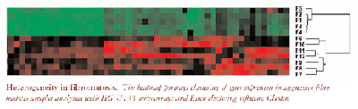
Amy Skubitz: For quality control, a pathologist looks at a frozen section at the time of surgery and then excess waste tissue is given to the staff of the Tissue Procurement Facility. The procurement facility staff makes their own quality control slide from tissue that is adjacent to what is later given to researchers. A pathologist will then verify the diagnosis, percent tumor, and percent necrosis on the H&E [hematoxylin and eosin] slide. We then select for those tissues that are greater than 90 percent tumor and less than 10 percent necrotic. Keith Skubitz: The microarray data that we've been publishing was generated by Gene Logic Inc. based on a contract with the University of Minnesota. Amy Skubitz: Each of the tissues that was provided to Gene Logic Inc. was excess; in other words, additional samples of the tissue were maintained at the University of Minnesota. For example, after pathology keeps the tissue that they need in order to make a diagnosis, the Tissue Procurement Facility may be provided with 10 grams of tissue, which they cut into multiple pieces. We use a piece of that tissue for immuno- histochemistry, while Gene Logic Inc. generates gene expression data with an adjacent piece of the tissue. So we have matching tissue that we can use to validate the gene expression data by immunohistochemistry or other types of experiments. Appropriate control selection
Obtaining healthy control tissue for many internal organ diseases is often a difficult task. The Skubitz’ weigh in on the challenges and solutions they’ve reached for collecting control tissue in ovarian and kidney cancer studies. Keith Skubitz: It may sound simple, but when selecting controls, the key thing is figuring out what question you want to answer. Without formulating the question, you can't really address anything else. For kidney cancer, we wanted to know if we could find therapeutic targets that would be expressed in the tumor, but would not be expressed in normal tissues. So in that case, the normal control is really the rest of the body. You could modify those conditions if you're willing to accept certain toxicity. Let's say you want the drug target expressed in kidney cancer, but not expressed in any essential tissue like heart or lung. In that case, you may not care if it's expressed in nonessential tissues—you don't need a thyroid, you don't need a prostate, you don't need an ovary for example. Amy Skubitz: We obtain the normal ovaries from women who come to the University of Minnesota for prophylactic oophorectomies. For example, women may require a hysterectomy or they may have a family history of some gynecological disease. When their ovaries are removed and the pathologist determines that the ovaries are normal, we can use them for our research projects. What complicates our studies is that the most common type of ovarian cancer arises from epithelial cells that are on the surface of the ovary. Therefore, it is difficult to do a direct correlation with surface epithelial cells from normal ovaries since the ovary may be handled by surgeons, residents, nurses, and pathologists. By the time the Tissue Procurement Facility staff gets the normal ovary, the surface epithelial cells may be rubbed off. When we eventually look at the gene expression data, it's with the understanding that we're comparing a sample of ovarian carcinoma, which has lots of cancerous epithelial cells in it, to a normal ovary, which may have a very small percentage of the normal counterpart of normal surface epithelial cells. That's why it is important for us to then do the immunohistochemistry and to see if the surface epithelial cells on normal ovaries stain for the proteins corresponding to the upregulated genes in the ovarian carcinoma samples. Furthermore, we've looked at over 300 other tissues, comprising tumors and normal tissues, to see whether the ovarian cancer expression signature would be differentially-regulated in them.  Statistics and software
Eisen Clustering and principle components analysis (PCA) are two major microarray analysis solutions frequently used by scientists. Each has strengths, but limitations as well.
Keith Skubitz: Eisen Clustering does have a nice visual effect, so it's very easy to readily understand what you are looking at. But it also has the obvious limitation that it's restricting the distribution to two-dimensional space. So PCA is definitely preferable, and that's what we've been using primarily, but we still do use Eisen Clustering as well. Amy Skubitz: One concern with PCA might be that you can rotate it any way you want in order to get clusters to appear. But, it's very satisfying when the clusters just leap right out at you. Keith Skubitz: PCA is usually preferable, but it is true that with either approach, depending on how you analyze the data, you can get different results. But these things we're manipulating are numbers, they're measurements, they're not magic. So we have to convince ourselves that whatever data or conclusion we're generating is actually true. One way to do that is by analyzing the data in several different ways and getting the same answer several different ways. One place where things can get confusing is in fold change analysis. A gene may be reported to be 50-fold up in group A versus group B. But before we would believe these results, we would like to actually look at a graphical representation of the data in each sample group, so that we can say, 'Yes, we believe this.' For example, it may be that 20 percent of the samples are contributing 90 percent of the signal, or something like that, but one would not be aware of that unless one looked at the individual data itself. Amy Skubitz: It's also important to look at the standard errors and p values for each of the data sets too. You can look at the actual data plotted out and see if there's a big spread or if they're all clustered at one end or the other. It's very time-consuming to do it that way, but if you don't, you can fall into this trap of believing that 50-fold up value. And then you end up choosing that gene without ever actually seeing the data. That's a concern. Common cancer signatures
Information from one type of cancer can help scientists learn about another. By comparing data, key genes and pathways can emerge that might otherwise be missed. Keith Skubitz: Right now we're focusing on specific tumors. One interesting point related to that question was a study we did looking for genes upregulated in leiomyosarcoma compared with normal tissues. We looked first at leiomyomas, which are benign smooth muscle tumors of the uterus, and we found some genes that were uniquely expressed compared with all the normal tissues. Later, we did an analysis of leiomyosarcomas, which are malignant tumors of smooth muscle. We looked for genes that were uniquely expressed in that sarcoma compared with a number of normal tissues, and we found some. But we then decided to go back and ask specifically, 'Were any of the genes that we found uniquely expressed in leiomyomas also selectively over-expressed in leiomyosarcomas compared with normal tissues?' In fact, this approach yielded new genes selectively expressed in leiomyosarcomas, but only a fraction of them. In other words, this approach identified heterogeneity in the leiomyosarcoma population, and allowed the identification of potential targets that were not found in our general screen. For example, we found that doublecortin was expressed in about half of the leiomyosarcomas. That's interesting, because we identified a subset of leiomyosarcomas and a potential drug target that we wouldn't have found without this clue from the earlier studies of related tumors. Experimental reproducibility
As the duration of their cancer research has grown, the Skubitz’ have run into the challenge of comparing data from different generations of sample collections and different generations of microarrays. The data are validating towards each other, but extra caution needs to be taken in these instances. Amy Skubitz: The experimental data that we initially analyzed was generated using the Human Genome U95 microarrays. It's just recently that we've used the Human Genome U133 chips and we've gone back to see if we could get some of the same data. Right now we're doing a different series of experiments with our ovarian cancer samples, and we're coming up with the same expression trends. Keith Skubitz: In kidney cancer, we're also finding a lot of the same genes with the U133 set that we found with the U95 set. So that's reassuring. Amy Skubitz: The HG U133 experiments are using the exact same tissue samples that were used in the HG U95 experiments. So it's not like taking a totally different tissue and seeing if we see the same trend. I would say that it's working well. Keith Skubitz: In many of the studies that we're doing we're looking at genes that are unique to the tumor versus a lot of different tissue types. I'd say it's been pretty reproducible. But let's say, for example, that you wanted to compare kidney cancer with normal kidney. The problem is that a kidney is not just a kidney. There's a cortex and a medulla; so depending on how much of your ‘normal kidney’ sample came from one part of the kidney versus another, the gene expression signal could be quite different. Also, in our first kidney cancer paper, we looked at gene expression in not only normal kidney and kidney cancer, but also in so-called diseased kidney. Not surprisingly, diseased kidney is not the same as normal kidney. So if one study looked at kidney cancer versus perfectly normal kidney, the results could vary depending on how much of the cortex or medulla were in the samples. Also, if there were inflammation in one of the kidneys, or it were somewhat atrophic, or they had significant hypertension or diabetes, or who knows what, the results could also differ. So, things that might at first appear to be the same comparison in different experiments, might not be. FOR MORE INFORMATION | Contact Information
![]() Amy Skubitz, PhD
Department of Laboratory Medicine and Pathology
University of Minnesota MMC #609
420 Delaware Street, S.E.
Minneapolis, MN 55455
Laboratory website ![]() Keith Skubitz, MD
Department of Medicine
508 Masonic Cancer Center
424 Harvard St. SE
Minneapolis, MN 55455
Laboratory website
Companies
![]() Affymetrix Inc.
![]() Gene Logic Inc. | Organizations:
![]() University of Minnesota Twin Cities
![]() International Society of Biological and Experimental Repositories Further Reading
![]() Skubitz KM, Cheng EY, Clohisy DR, Thompson RC, Skubitz AP. Differential gene expression in liposarcoma, lipoma, and adipose tissue. Cancer Invest 2005; 23(2):105-18.
![]() Skubitz KM, Cheng EY, Clohisy DR, Thompson RC, Skubitz AP. Gene expression in giant-cell tumors. J Lab Clin Med 2004;144(4):193-200.
![]() Skubitz KM, Skubitz AP. Characterization of sarcomas by means of gene expression. J Lab Clin Med 2004; 144(2):78-91. | ![]() Skubitz, KM, and Skubitz, APN. Differential gene expression in renal cell cancer. J Lab Clin Med 2002; 140:52-64.
![]() Hibbs, K, Skubitz, KM, Pambuccian, SE, Casey, RC, Burleson, KM, Oegema, TR, Jr., Thiele, JJ, Grindle, SM, Bliss, RL, Skubitz, AP. Differential gene expression in ovarian carcinoma: identification of potential bio-markers. Am J Pathol 2004; 165(2):397-414.
![]() International Society of Biological and Experimental Repositories (ISBER). Best Practices for Repositories I: Collection, Storage, and Retrieval of Human Biological Materials for Research. Cell Preservation Technology 2005; 3(1):5-48.
Tools for download
Eisen Clustering Software |
|


